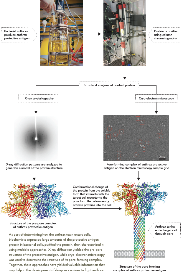
Anthrax disease is caused by the bacterium Bacillus anthracis. Anthrax toxin consists of three components, one of which is a protein called protective antigen. To study how the protective antigen works, the protein was produced in bacterial cells, then purified using column chromatography. Crystallization and X-ray diffraction analysis yielded the structure of the soluble complex that interacts with the target cell’s receptors before the protein is inserted in the cell membrane. More recently, by working at dilute conditions directly on the electron microscopy sample grid, isolated complexes of the protective antigen in the pore-forming conformation were obtained, enabling its structure to be determined by cryo-electron microscopy. Together these studies and others have provided insight into the multistep process by which this membrane-spanning channel is formed, and this information can be used to design protective measures to prevent the entry of anthrax toxin proteins into cells.